A Research Infrastructure
The core technology at CERM/CIRMMP is NMR spectroscopy, and the onsite instrumentation is among the most advanced in the world. A European transnational access service, funded by EC since 1994 in addition to service provision at national level operating since 1990, places CERM/CIRMMP at the top of the list for experience among the European NMR Research Infrastructures.
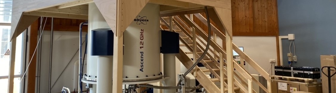
The flagship of the Center is the impressive collection of NMR spectrometers which feature the largest magnetic field range in the world (from 1200 MHz to the earth field) and ranks it among the best equipped laboratories in the world. The NMR labs are flanked by molecular biology laboratories where samples for the NMR are produced.
Solution and Solid State NMR Spectrometers
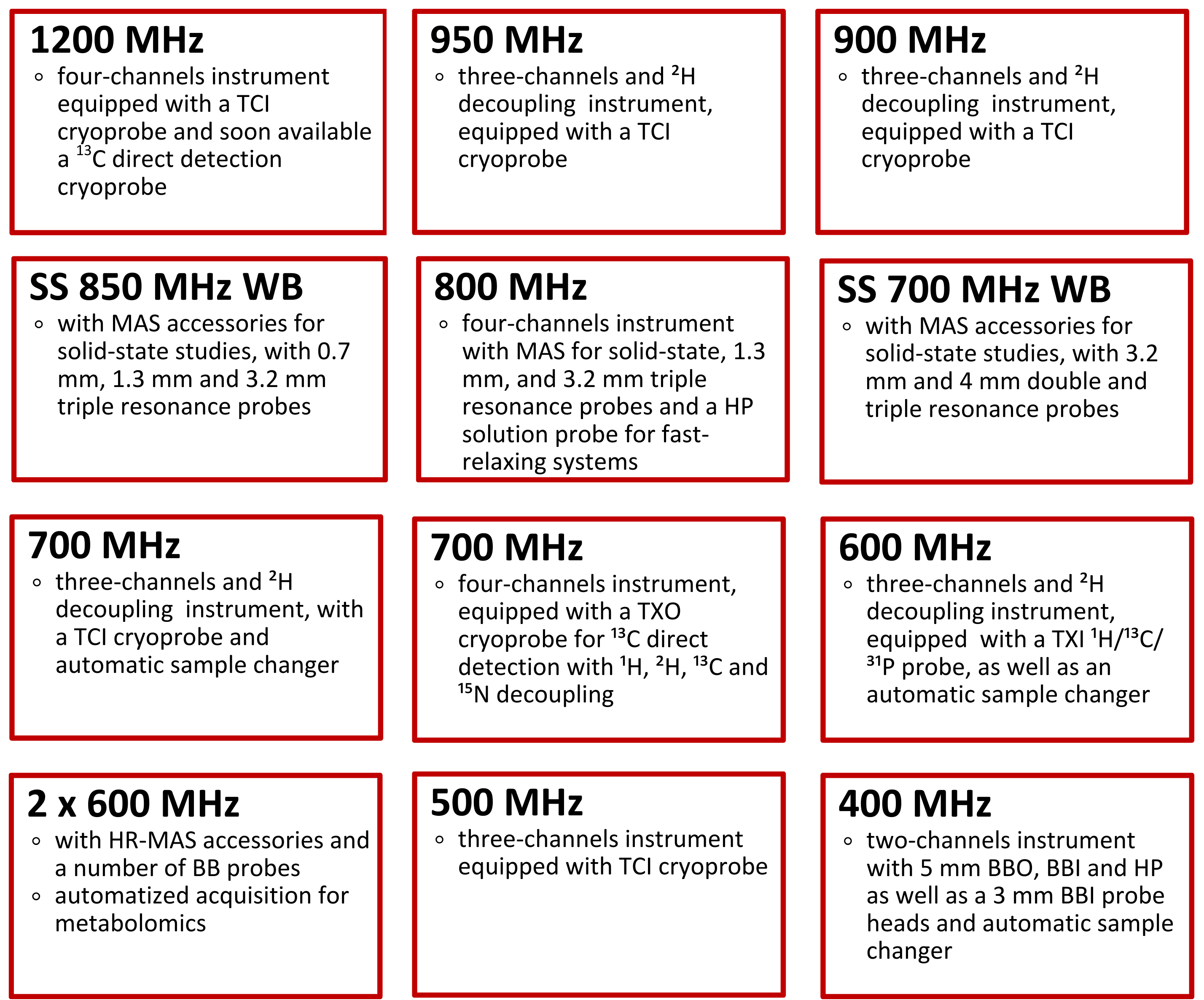 All NMR instruments are state-of-the-art, digital spectrometers equipped with a variety of cryoprobes as well as of specific probes covering a broad range of frequencies and of observable nuclei.
In addition to all the standard pulse sequences for spectroscopic, structural, dynamical, and functional characterisation, tailored pulse sequences for structural determination of high molecular weight proteins and paramagnetic systems are implemented, as well as 13C direct-detection solution protocols for “protonless” NMR experiments and structural characterisation of biomolecules, including unfolded or partially unfolded ones. Pulse sequences and experiment setup have been implemented for the detection and characterisation of paramagnetic systems and in this field CERM has been pioneer since decades.
A detailed list of the instrumentation is available here
EPR
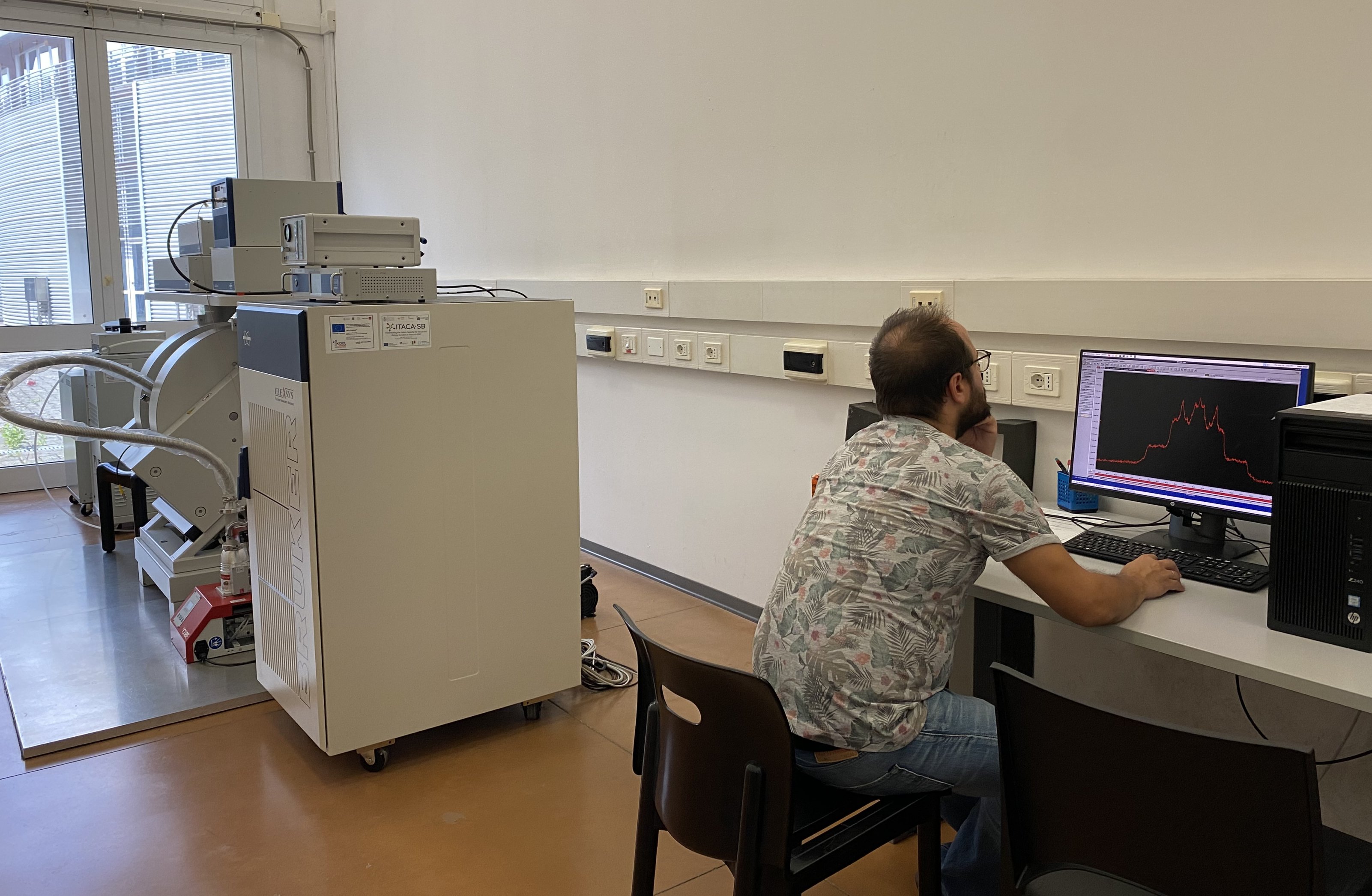 CERM has installed a Bruker ELEXYS E580 spectrometer operating at X-band (9.8 GHz) and Q-band (34 GHz).
The EPR spectrometer at the X-band is equipped with a SHQE resonator and an Oxford ESR900 cryostat. Continuous wave measurements, such as paramagnetic metals characterization and site-directed spin labelling applications, can be performed from room temperature to 4K.
The EPR spectrometer at Q-band is equipped with an EN5107D2 resonator, an Oxford CF935 cryostat and a 3W-AWG microwaves amplifier.
Different pulse-EPR experiments can be carried out:
- Spin echo techniques to measure electron spin relaxation times
- ESEEM and 2D-HYSCORE experiments for the analysis of hyperfine interactions between electron and nuclear spins to obtain information about spin density and distances
- DEER experiments to measure long range distances with electron-electron dipolar coupling.
The spectrometer is equipped with a newly installed DICE-II Pulse ENDOR System E560D-P-RF for spectroscopic characterization of the molecular and electronic structure of paramagnetic species.
Fast Field Cycling (FFC) relaxometer
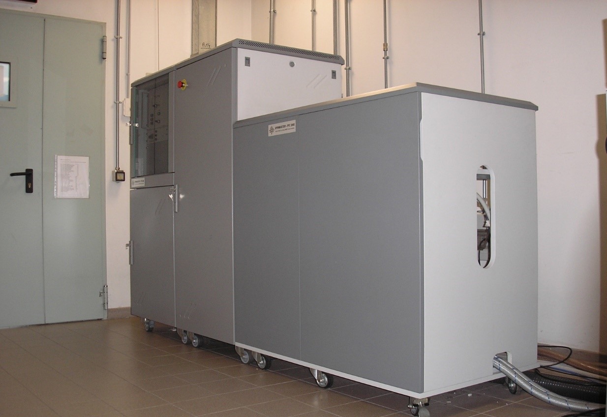 The Fast Field Cycling (FFC) relaxometer allows for the measurement of the nuclear spin-lattice relaxation rate constants as a function of the applied magnetic field, from 0.01 MHz to 40 MHz proton Larmor frequency. Due to the lack of resolution, experiments are usually performed by measuring the relaxation rate of bulk water protons in solutions with diamagentic macromolecules or paramagnetic complexes. The colleced profiles provide direct information of the spectral density functions of the detected nuclei, and thus on the strength of the interactions and on the dynamics of molecules over a wide frequency range. This technique is largely used for the characterization of candidate contrast agents for MRI.
Computational Facility
 CERM/CIRMMP provides integrated databases and software for genome browsing, metal binding analysis, structure calculation with/without paramagnetic restraints, sequence valida-tion, domain organisation, evolution, protein complex analysis. Access to programs for NMR data processing and structural calculations is also provided via web.
CERM/CIRMMP maintains a node of the European Grid Initiative. The available hardware comprises two clusters with 80 and 1024 CPU-cores respectively, and four TB of shared stor-age. A cluster with six Nvidia Tesla K20 GPGPU cards is also available.
Biological and Biophysical Equipment and Facilities
 CERM/CIRMMP is equipped with state-of-the-art facilities for gene cloning and protein ex-pression and purification. Large scale protein expression in prokaryotes and yeast is available through the use of fermenters. Different isotope labelling schemes, including specific labelling schemes oriented to NMR characterisation, can be achieved through the use of auxotrophic strains. Fully equipped facilities for protein purification are available, including last-generation instruments for streamlined purification (ÄKTA pure chromatography system) and equipment for protein purification and reconstitution in anaerobic environment (glove box). A mammalian expression lab for in-cell NMR is also available.
Multi Angle/Dynamic Light Scattering - Instrument for measurements on batch samples or on in-flow samples (FPLC coupling).
Isothermal Calorimetry (ITC) - ITC device to measure thermodynamical parameters in microsamples. The instrument is fully equipped for studying protein-ligand and protein-protein thermodynamical parameters.
Optical Spectroscopy - Absorption/Fluorescence Spectrophotometer operating from 1000 to 200 nm, Circular Dichroism (CD) spectrometer operating form 1200 to 200 nm (Near-IR, Visible, UV) to derive information on the proteins secondary structure or protein-metal interaction, and stopped-flow spectrophotometer are available in the infrastructure.
Optical Microscope (Nikon ECLIPSE TsR-FL) - Compact inverted fluorescence microscope, perfect for small labs without compromising on performance.
Flow Cytometer (Guava® easyCyte™) - designed to simplify flow cytometry with its novel microcapillary technology in a compact, user-friendly format
SEC-MALS (Dawn 18 System) - perfect for analyzing macromolecules and nanoparticles, it measures hydrodynamic radius and separates particles by size.
Surface Plasmon Resonance - SPR device for studying protein-ligand or protein-protein interactions
Circular Dichroism - CD device to gain information on the secondary structure of proteins
Fluorimetry (Varian Eclipse) - fluorimeter for 200-900 nm measurement equipped with a xenon flash lamp that allows fluorescence measurements between 200 to 900 nm. It offers various accessories for temperature control, surface analysis, and high-throughput measurement.
Stopped-flow Spectrophotometer
Glove box
Cryo-EM
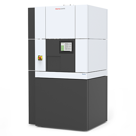 Cryo-Electron Microscopy (Cryo-EM) applies Transmission electron microscopy (TEM) to image particles, including macromolecular complexes, viruses, and cells in their native state at cryogenic temperatures.
Recent technological advances in sample preparation/vitrification strategies, computation and especially instrumentation (new image processing algorithms, Direct Electron Detection camera, Volta phase plate) allowed Cryo-EM single particle analysis (Cryo-EM SPA) to become a newly dominant discipline, accompanied by X-ray crystallography and nuclear magnetic resonance (NMR) spectroscopy. It has prompted revolution in structural biology, allowing researchers to use Cryo-EM to solve near-atomic-resolution macromolecular structures.
In Cryo-EM SPA, three-dimensional density maps can be reconstructed by averaging a large number of two-dimensional projection images of the same object along different directions. It covers a wide molecular weight range of specimens, from protein complexes in the tens of kilo Daltons to large virus particles with hundreds of mega Daltons. Moreover regular fibrillar protein aggregates (amyloids) have also been successfully targeted through cryo EM, further highlighting the impact of this approach on biological and biomedical research.
CERM/CIRMMP has full access to the Inter-departmental Florence Center for Electron Nanoscopy (FloCEN), that houses state-of-the-art equipment for cryo-EM. This includes a Glacios (Thermo Fisher Scientific) 200-kV, also equipped with a Falcon III direction electron detector, and a Vitrobot (Thermo Fisher Scientific) specimen preparation unit that completely automates the vitrification process.
X-ray Crystallography
CERM/CIRMMP is equipped with standard crystallisation facilities and with an automated nano-dispensing device (Mosquito, TTP Labtech). Furthermore it has full access to the Inter-departmental Crystallography Centre of the University of Florence (CRIST) equipped, among other instruments, with a sealed-tube diffractometer. The most recent one is a Bruker D8 Venture with double microsource (Cu and Mo) bearing a Photon III Pixel Array detector and the older one is an Xcalibur PX Ultra (Oxford Diffraction) equipped with a 165 mm CCD detector for routine in-house data collections. Both diffractometers are equipped with a liquid nitrogen cryosystem. Regular access to synchrotron beam time slots in European facilities is also available.